


You are here
Tumor Pathology Department
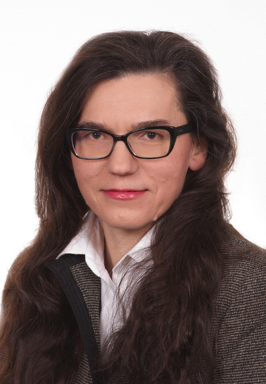
Head
dr hab. n. med. Ewa Chmielik, MD
Secretariat:
telephone: +48 32 278 94 01, fax: +48 32 278 94 15
Medical secretariat:
telephone: +48 32 278 94 02, +48 32 278 94 70, +48 32 278 94 65.
The Maria Sklodowska-Curie Memorial Institute Tumor Pathology Department, Gliwice ranks high as one of the most modern and best equipped pathology departments in Europe thanks to the steady implementation of current knowledge and techniques in the examination process.
Our department provides a leadership role in residency training and continuing education in pathology, cytology, and oncology. Our pathologists actively participate in the most prestigious world congresses, meetings, seminars, and training courses. The Department employs highly specialized physicians and experienced laboratory technicians. The Pathology Department has been performing fine needle biopsies for over 25 years now. At present, we do about 2,000 biopsies per month, a fact that positions us among centers with the highest number of fine needle biopsies in Europe.
Our Pathology Department has been performing gynecological examinations since the day it was first established in 1947. We were among the first pioneers to introduce the smear evaluation method according to the Bethesda system. Microscopic examinations are performed using the best available microscopes equipped with an additional consultation arm.
In our diagnostic work, we always give careful consideration to the clinical data comprised in the patient’s case history.
We are the second pathology department that is prepared to apply telepathology. We will soon be able to start Internet-based slides consultations with the best international pathology centers.
The Department boasts a state-of-the-art fully automated cryostat processing frozen sections of all tissue material.
The Immunohistochemistry Laboratory of the Tumor Pathology Department was established in 1993 as one of the first in the country. Since then, it has led the field in a wide spectrum of immunohistochemical testing.
PERFORMED EXAMINATIONS
We perform lung biopsies under continuous X-ray monitoring.
Cytologic material undergoes complex evaluation applying immunohistochemistry flow cytometry and digital image analyses. Depending on the requirements, the cytologic material is assessed for:
Gynecologic Cytology
We specialize in assessing smears sampled after radiotherapy. We introduce in situ hybridization and the PCR method for detecting oncogenic HPV viruses.
Histologic Diagnosis
The Tumor Pathology Department is equipped with the latest apparatus for preparing histopathologic slides. The fully automated tissue processing, staining, and covering allows for full standardization of quality. When required, tissue material is processed by applying special techniques: histochemistry and immunohistochemistry. We specialize in trepanobioptats evaluation in cases of lymphatic system neoplastic proliferation.
Frozen Section (Intraoperative Examination)
The high quality of frozen sections (comparable to paraffin testing) not only allows for determining the presence and nature of a lesion but also to define the adequacy of surgical margins during surgery, especially in the case of breast conserving and reconstructing surgery. At present, we carry out about 600 frozen sections per month, a part of which is ordered by hospitals from the regions of Silesia and Opole, which co-operate with us on a regular basis.
Immunohistochemical Testing
Our Department ranks among the top laboratories in this country, a fact underpinned by the positive findings of the International Quality Control NordiQC. We are fully equipped with staining platforms, both Dako Autostainer Link and Ventana BenchMark Ultra. We have a wide range of antibodies which we continuously enlarge so as to guarantee our patients the most precise differential diagnosis.
Flow Cytometry
The following tests are carried out by the Flow Cytometry Lab:
Consulting
USG guided fine needle biopsies of the:
- Thyroid
- Breast tumors
- Abdominal cavity organs
- Lymph nodes
- Soft tissue tumors
- Integuments
- Salivary glands
- etc.
We perform lung biopsies under continuous X-ray monitoring.
Cytologic material undergoes complex evaluation applying immunohistochemistry flow cytometry and digital image analyses. Depending on the requirements, the cytologic material is assessed for:
- presence of a particular antigen - differential diagnostics of neoplasms,
- presence of estrogen and progesterone receptors,
- roliferating activity of tumor cells and DNA ploidy using flow or static cytometry.
Gynecologic Cytology
We specialize in assessing smears sampled after radiotherapy. We introduce in situ hybridization and the PCR method for detecting oncogenic HPV viruses.
Histologic Diagnosis
The Tumor Pathology Department is equipped with the latest apparatus for preparing histopathologic slides. The fully automated tissue processing, staining, and covering allows for full standardization of quality. When required, tissue material is processed by applying special techniques: histochemistry and immunohistochemistry. We specialize in trepanobioptats evaluation in cases of lymphatic system neoplastic proliferation.
Frozen Section (Intraoperative Examination)
The high quality of frozen sections (comparable to paraffin testing) not only allows for determining the presence and nature of a lesion but also to define the adequacy of surgical margins during surgery, especially in the case of breast conserving and reconstructing surgery. At present, we carry out about 600 frozen sections per month, a part of which is ordered by hospitals from the regions of Silesia and Opole, which co-operate with us on a regular basis.
Immunohistochemical Testing
Our Department ranks among the top laboratories in this country, a fact underpinned by the positive findings of the International Quality Control NordiQC. We are fully equipped with staining platforms, both Dako Autostainer Link and Ventana BenchMark Ultra. We have a wide range of antibodies which we continuously enlarge so as to guarantee our patients the most precise differential diagnosis.
Flow Cytometry
The following tests are carried out by the Flow Cytometry Lab:
- Phenotyping of blood and lymph glands (evaluation of lymphocytes T, B and subpopulation of cellular proliferative diseases of the hematopoietic system);
- Before autologous and allogenic transplantations, both hematopoietic progenitor CD34+ cells and dead cells obtained during separation are evaluated quantitatively in the donors’ stem or blood;
- Determination of DNA ploidy of malignant cells in primary tumors, that is evaluation of the tumor proliferating activity;
- Processes involved in the cell death;
- Suppressor genes of tumors;
- In vitro effects of drugs.
Comparing the results obtained during the phenotypic analysis using a flow cytometer together with morphological and histological evaluation as well as genetic testing allows for a detailed analysis of the diseases of the hematopoietic system.
Molecular Methods
Molecular Methods
Our department was among the first research facilities in Poland to implement fluorescence in-situ hybridization (FISH) for routine clinical evaluation of the HER-2 gene status, which is currently a necessary component of selecting an individual therapy for patients with breast and stomach cancer (personalized medicine). The Department operates a FISH external quality control program and successfully passes the audit each year. We also use the FISH method for differential diagnosis of soft tissue tumors, lymphomas, as well as head and neck tumors. We apply a wide range of diagnostic methods for the detection of oncogenic human papillomaviruses (HPV). As the only oncology center in Poland so far, we have implemented a unique, certified test to detect the presence of 14 HPV types based on a quantitative PCR method. The most advanced PCR technique helps us achieve – in the opinion of the Clinic - reliable and valuable results in the prevention of cervical cancer and individualized treatment of oropharynx cancer patients. Furthermore, in the case of HPV infections, by using the imaging techniques we are able to identify chromogenic in-situ hybridization (CISH) and immunohistochemistry in the pathologic material. In the near future, the Department will launch one of the most modern systems for gene structure analysis (Next Generation Sequencing) that will allow for a two-day testing of the 200 genes associated with malignancy, based on the patient’s standard histological material. This system in conjunction with a scanner microscope slides and macrodissection device and create a matrix of tissue allow the analysis of mutations and other genetic disorders in specific, well-defined fragments of tumor.
Consulting
The Oncology Center Tumor Pathology Department provides consultation services in approximately 3,500 histopathological and cytological cases per year, with samples sent in from all over the country. Our staff have extensive experience in thyroid pathology, endocrine pathology, neuropathology, head and neck tumors, breast pathology, gastrointestinal pathology, hematopathology, pulmonary pathology, gynecopathology, male genitourinary pathology, renal tumors, soft tissue and bone tumors, or rare tumors. As a well-known Polish Reference Center for Thyroid Carcinoma, we consult in over 1,000 cases of thyroid tumors per year as well as offer immunohistochemical diagnostics.
 Polski