


You are here
Radiology and Diagnostic Imaging Department
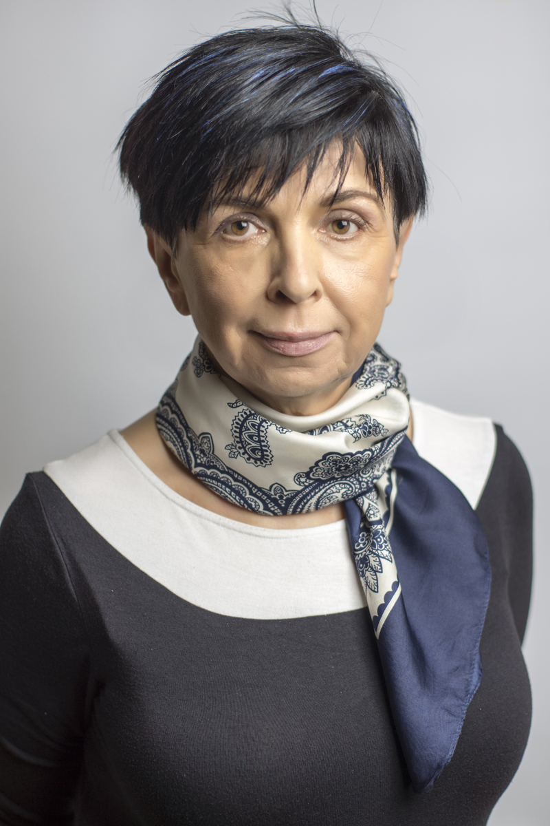
Head
prof. Barbara Bobek-Billewicz, PhD, MD
Secretariat
telephone: +48 32 278 93 65, fax +48 32 278-93-59
e-mail: rtg_sekretariat@io.gliwice.pl
Registration Desk
telephone: +48 32 278 93 58
Early Breast Cancer Detection Program
- Computed axial tomography (CAT scan),
- Magnetic resonance imaging (MRI scan): classical MR imaging of all anatomical areas and the entire body, blood vessel testing and functional imaging (spectroscopy, diffusion imaging, perfusion, brain mapping),
- Classical radiology (digital tests),
- Digital mammography,
- Galactography,
- Ultrasonography (Color and Power Doppler, endocavitary, elastography, Automated Breast Volume Scanner [ABVS]),
- Fine-needle biopsy using different imaging techniques,
- Thick-needle biopsy and thick-needle biopsy with ultrasound, CT, and MRI tests.
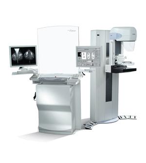
Three platforms are used to perform mammography tests, i.e. GE Seno Pristina, Siemens Mammomat Inspiration Prime (Progressive Reconstruction Intelligently Minimizing Exposure) with True 3D Breast Tomosynthesis, and Hologic Selenia 2, or on a Mammobus (Siemens Mammomat Inspiration). There is also another option to use breast USG and MRI as a further diagnostic procedure. Our Computer Aided Detection (CAD) automatically detects breast lesions.

The Radiodiagnostics Department is equipped with two digital platforms: Arcoma Precision i5 and Siemens AXIOM Luminos dRF, with a flat detector for conventional diagnostics of the chest, abdominal cavity, digestive and urinary systems, and bones, thereby guaranteeing the highest imaging quality.
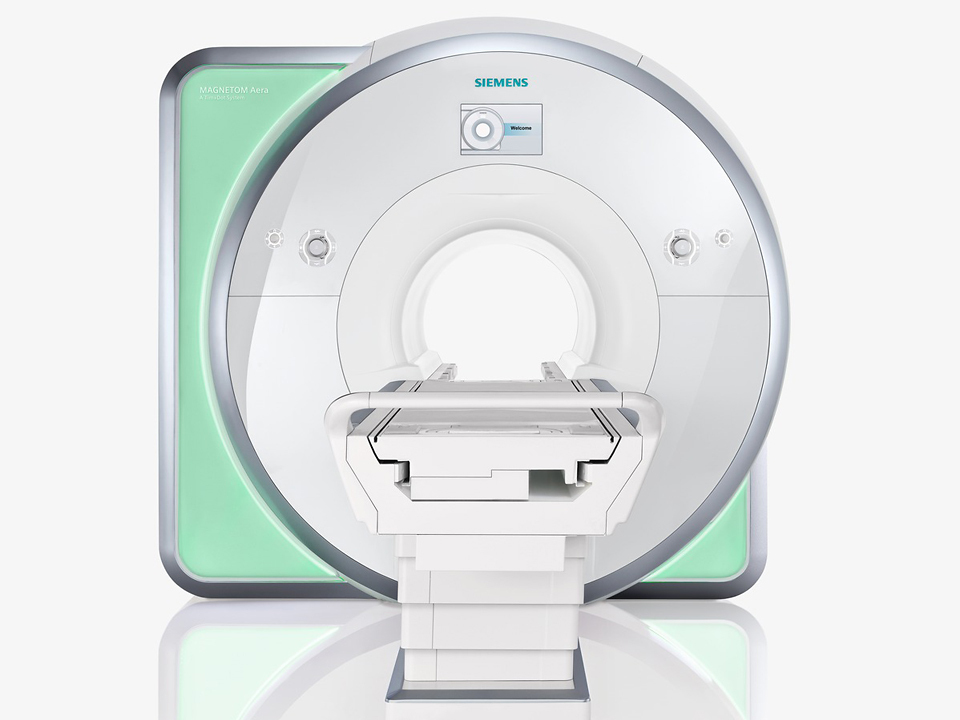
The MRI department is equipped with three modern MR platforms, i.e. Siemens Magnetom Sola Fit 1,5 T and Prisma and Vida 3T (the only such platform in the Upper Silesian Province to feature a 3T magnetic field induction). The MR tests range encompasses the following: classical MRI, MR angiography, MR cholangiography, and advanced techniques, such as diffusion-weighted imaging (DWI), perfusion-weighted imaging (PWI), spectroscopy (MRS), functional magnetic resonance imaging (fMRI BOLD), diffusion tensor imaging of cerebral white matter (DTI), and whole body MR. Prostate exams using spectroscopy are performed with a surface coil. For breast examination (MR mammography), apart from classical imaging, we also perform diffusion imaging and spectroscopy testing. Brain spectroscopy results are additionally analyzed by an LC model professional software. The intravenous contrast agent is administered using a two-channel automated syringe. Within the framework of cooperation with the Silesian Center for Heart Diseases in Zabrze, we perform a comprehensive range of heart MR testing.
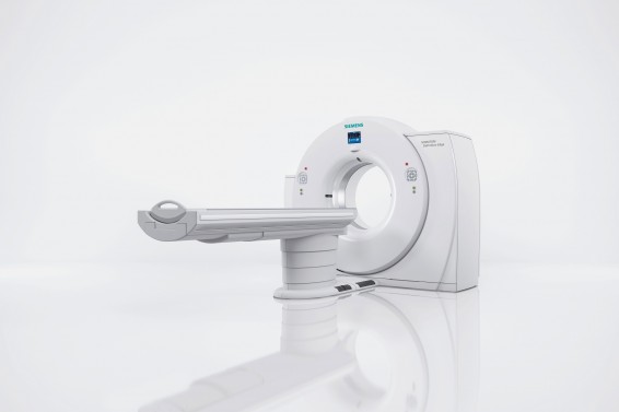
The Department has two multi-row computed tomographs (CT), i.e. Siemens Definition Edge Plus (128 layers) and Philips IQon CT (one of a kind dual energy device in Poland). The machines are designed for patient examinations using both spiral and sequence techniques. We perform densitometry tests of all the organs and anatomical areas (except for the heart). A special program helps detect tumor lesions in lungs and to analyze the image over time. To minimize the risk of complications, we use modern, non-ion, low-osmolar contrast agents. In order to obtain the best possible image, the contrast agent is administered by a two-channel automated syringe.
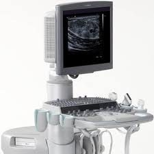
Ultrasonography Department
The Department has five modern USG platforms with Color Doppler and Color Power Doppler options. Three of them are suitable for performing elastography of lesions in breast glands, and one of them - in the liver. In the Ultrasonography Department, we examine the neck, the abdominopelvic cavity, and the breasts.
 Polski